FACILITY AT NFFA-TRIESTE
THE SOURCE
The new SPRINT (Spin Polarized Research Instrument in the Nanoscale and Time Laboratory) has been equipped with two laser sources (PHAROS) with a common oscillator, both emitting pulses at 1030 nm, with time duration of 300 fs, variable repetition rate from 50 kHz up to 1 MHz, pulse energy of 400 µJ and average power up to 20 W. These lasers are at the heart of the new HHG-based beamline.
As in fig. 2, the output pulse from the laser at 1030 nm is splitted by a variable beam splitter, in order to allow a proper pump-probe setup with two spatially and temporally overlapped beams to be implemented. The power ratio for our optical system is 90% reflected and 10% transmitted. Each of the two beams can be used for either pumping or probing, depending on what the experiment requires. As an example, the two beam profiles at 1030 nm and 515 nm are reported in fig. 3.
One of the beams is sent on a beta barium borate (BBO) crystal of about 2 mm thickness, which produces the 2nd harmonic at 515 nm with 50% efficiency; two wavelength separators serve the purpose of removing the fundamental while keeping the 2nd harmonic in the optical path. After a series of mirrors, the beam enters the generation chamber, where one 10 cm focal length lens produces a 40 µm Gaussian beam that allows to reach the 1014 W/cm2 peak power density.
In the generation chamber the generation gas is inserted by means of a short and narrow (70 mm internal diameter) capillary nozzle. The whole interaction region is pumped via an in-vacuum high diameter tube feedthrough, and the chamber itself is pumped by a magnetically levitated pump and a primary scroll. The pumping system has been designed to allow operation even in high backing pressure of the nozzle (up to 10 bars) and to reduce as much as possible the pressure near the laser focus to avoid X-ray absorption.
The beam emitted by the gas is composed of a superposition of the odd harmonics of the fundamental frequency of the laser. The water-refrigerated pinhole connecting the generation and the monochromator chambers blocks the 515 nm fundamental radiation from propagating further, as well as functioning as a vacuum impedance. The single-grating monochromators mounted in the off-plane geometry serve the purpose of selecting one specific harmonic and conserving the time duration of the pulse. The harmonics are dispersed in the direction perpendicular to the beam propagation plane; by rotating the grating along the optical axis of the system, the desired spectral range is reflected on a second toroidal mirror, which focuses horizontally the radiation.
The selected harmonic may be measured in intensity in a dedicated chamber, where a channeltron can be inserted in the beam path and the photocurrent measured. If the channeltron is removed, the harmonic can propagate until meeting a refocusing mirror that compensates the horizontal divergence due to focussing on the slits. The beam then grazes the edge of the beam alignment mirror and propagates onto the sample in the endstation.
Other than seeding the HHG process, the lasers may power three Optical Parametric Amplifiers (OPAs). These systems generate the second harmonic of the fundamental with high efficiency, produce white light in a non-linear crystal and amplify a selectable wavelength of white light: the process results in an almost continuous (5 nm steps) tunable wavelength conversion with more than 10% efficiency. The two light conversion ORPHEUS HP and ORPHEUS ONE HP cover respectively the 210-2500 nm and 1350-16000 nm ranges with pulse lengths of 200 fs, while the newly installed ORPHEUS F covers the 630-2500 nm range with a pulse length between 30 and 50 fs. The beams are then sent in a delay line, which allows a fine tuning of the temporal distance between pump and probe pulses: this means that the signal of the photoelectrons in the detectors can be related to a specific instant in time.

IMAGE PREVIEW
Fig. 1: Picture of the experimental chamber during the commissioning with a pulsed electron beam.
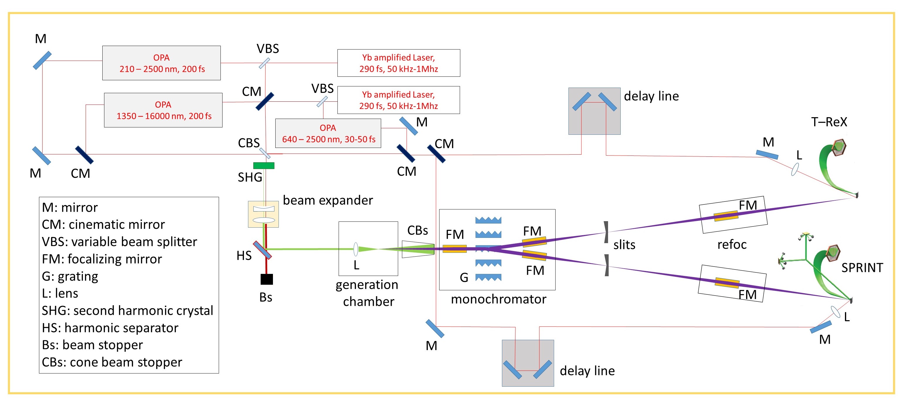
IMAGE PREVIEW
Fig. 2: Pump-probe setup.
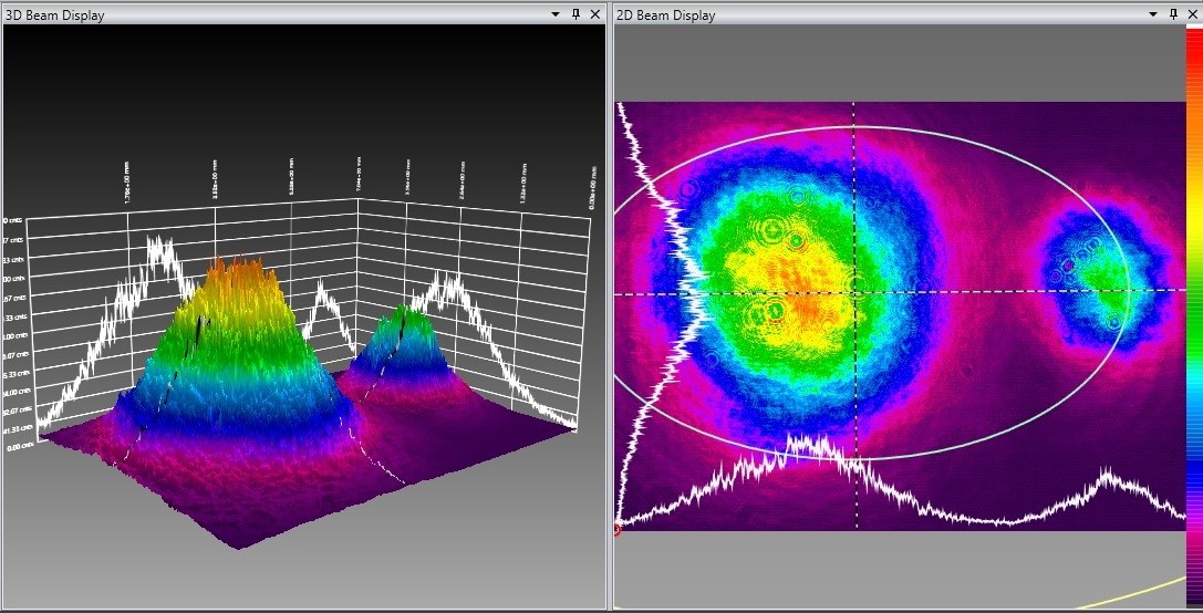
IMAGE PREVIEW
Fig. 3: 3D (left panel) and 2d (right panel) profile of 1030 nm and 515 nm after the prism separation. The higher intensity peak is at 1030 nm.
THE METHOD

IMAGE PREVIEW
Fig. 4: Image of the endstation. The two UHV chambers are independent and can operate simultaneously. Here, thin films were being grown in the preparation chamber while other samples were measured in the main chamber.

IMAGE PREVIEW
Fig. 5: A 3D rendering of the endstation. A sagittal section of the main chamber exposes the electrostatic lenses that transport the photoemitted electrons to the detectors and the two orthogonal scattering chambers (yellow).
The main instrument is a Mott detector measuring the spin-polarization of the electrons photoemitted under the photon pulse excitation.
When using the HHG source, the spin-polarization signal comes mostly from the secondary electrons, which undergo several inelastic collisions when travelling in the material. For this reason, a high spin-polarization is observed in the extremely low energy component of the photoelectron spectrum; this value is proportional to the magnetization state of the sample. Such information is retrieved only from the extreme surface, as experiments of overlayer deposition proved that the magnetization signal is lost after deposition of few Å of non-magnetic material.
Conversely, the low-energy photons provided by the OPAs allow to emit electrons from the near-Fermi region with minimal kinetic energy: this threshold photoemission regime is characterized by no average magnetization signal, but gives access to the magnetic moment of the electrons in the bands. The low kinetic energy of the photoelectron means also that the probing depth exceeds several layers of material, making it a more bulk-sensitive technique.

IMAGE PREVIEW
Hysteresis measured on the horizontal polarization channel in continuous mode on a test thin film Fe/Si (001) sample. The curve was acquired under a continuous excitation and completed in less than 15 minutes. The reduced signal-noise ratio at larger fields is due to the drop of intensity produced by the Lorentz deflection by the external field of the magnet
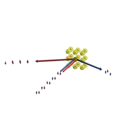
IMAGE PREVIEW
A simplified picture of Mott scattering process, in which spin-orbit interaction in elastic high energy scattering from gold atoms partially selects the electron trajectories on the basis of their spin state.
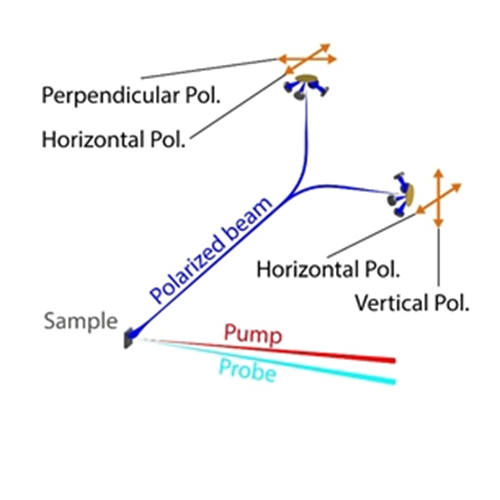
IMAGE PREVIEW
A simplified scheme of the experimental geometry: the pump laser beam drives the material out of equilibrium, and the probe UV or X-ray pulse generates photoemission. The polarized electron beam is sent alternately to the two targets so that the full polarization vector is reconstructed.
The spin-polarization of the photoemitted electrons is measured through the classical Mott scattering asymmetry: electrons accelerated to 40keV hit a gold target and are then detected by two detectors at ±120° in the scattering plane. Spin-orbit interaction with gold nuclei results in an asymmetry dependent on the polarization of the incoming beam. Such asymmetry is then proportional to the polarization of the incoming electron beam along the direction perpendicular to the scattering plane:

A vectorial Mott detector features two orthogonal scattering chambers in which eight detectors are mounted, featuring a total of four scattering planes: three are orthogonal and one is redundant and used to cross calibrate the signals. An electrostatic switch allows to change in no time from one target to the other, realizing a quasi-simultaneous measurement of the three independent components of the spin-polarization.
The instrument has been mounted in a dedicated UHV chamber, where a suitable sample environment has been designed to meet the demanding criteria of such kind of measurements. The complex signal readout was upgraded with high-performance electronics able to operate both in the pulsed, high peak flux regime (for pump-probe experiment with ultrashort pulses) and in the continuous steady flux mode (the standard mode for synchrotron or laboratory sources).
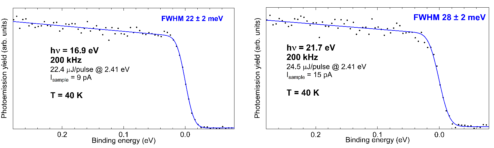
IMAGE PREVIEW
Fig. 6: Fermi edges at 16.9 eV and 21.7 eV measured at 200 kHz repetition rate. The curve fit retrieves the harmonic energy bandwidth.
On top of the spin-polarization analysis, the endstation is equipped with a hemispherical electron analyser (SCIENTA 2002, r = 200 mm) with less than 1 x Ep resolution. The SCIENTA analyser is the main tool for the investigation of the energy resolution of the HHG pulse, which has been measured by means of the acquisition of the Fermi edge of polycrystalline Au at low temperature. Fig. 6 shows the spectra acquired with the 7th and the 9th harmonic and their correspondent overall energy resolution. The energy bandwidth of 22 ± 2 eV combined with the measured temporal resolution of 105 ± 45 fs means that the radiation is near the transform limit, which in turn allows to measure superb quality valence band photoemission spectra.

IMAGE PREVIEW
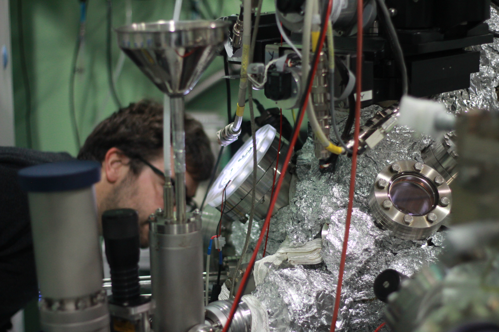
IMAGE PREVIEW

IMAGE PREVIEW

IMAGE PREVIEW
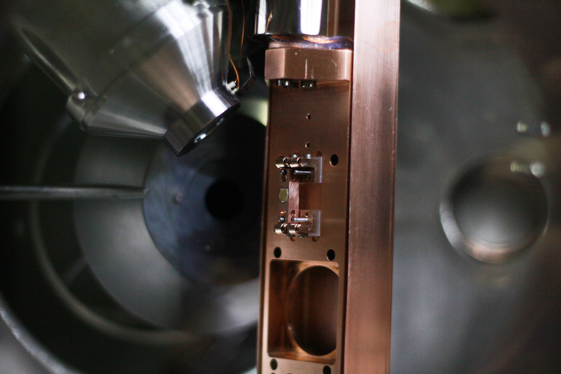
IMAGE PREVIEW

IMAGE PREVIEW
