Infrared scattering-type scanning near-field optical microscopy (IR s-SNOM) and imaging is here exploited together with attenuated total reflection (ATR) IR imaging and scanning electron microscopy (SEM) to depict the chemical composition of fibers in hybrid electrospun meshes. The focus is on a recently developed bio-hybrid material for vascular tissue engineering applications, named Silkothane®, obtained in the form of nanofibrous matrices from the processing of a silk fibroin-polyurethane (SFPU) blend via electrospinning. Morphology and chemistry of single fibers, at both surface and subsurface level, have been successfully characterized with nanoscale resolution, taking advantage of the IR s-SNOM capability to portray the nanoscale depth profile of this modern material working at diverse harmonics of the signal. The applied methodology allowed to describe the superficial characteristics of the mesh up to a depth of about 100 nm, showing that SF and PU do not tend to co-aggregate to form hybrid fibers, at least at the length scale of hundreds of nanometers, and that subdomains other than the fibrillar ones can be present. More generally, in the present contribution, the depth profiling capabilities of IR s-SNOM, so far theoretically predicted and experimentally proven only on model systems, have been corroborated on a real material in its natural conditions with respect to production, opening the room for the exploitation of IR s-SNOM as valuable technique to support the production and the engineering of nanostructured materials by the precise understanding of their chemistry at the interface with the environment.
It is well-known that all the phases of the manufacturing influence the extraordinary aesthetic and acoustic features of Stradivari’s instruments. However, these masterpieces still keep some of their secrets hidden by the lack of documentary evidence. In particular, there is not a general consensus on the use of a protein-based ground coating directly spread on the wood surface by the Cremonese Master. The present work demonstrates that infrared scattering-type scanning near-fields optical microscopy (s-SNOM) may provide unprecedented information on very complex cross-sectioned microsamples collected from two of Stradivari’s violins, nanoresolved chemical sensitivity being the turning point for detecting minute traces of a specific compound, namely proteins, hidden by the matrix when macro or micro sampling approaches are exploited. This nanoresolved chemical-sensitive technique contributed new and robust evidence to the long-debated question about the use of proteinaceous materials by Stradivari.
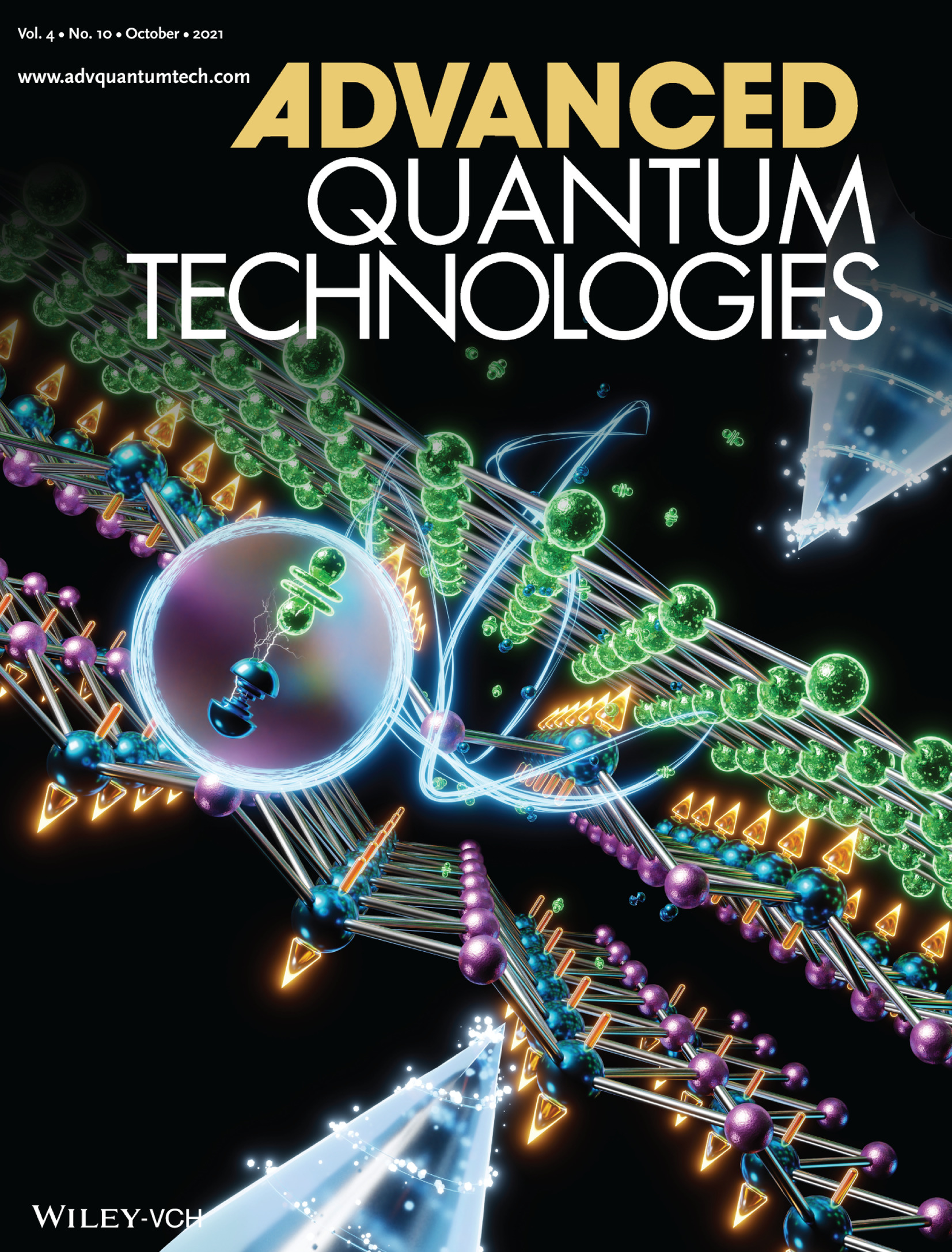
Topological materials are a promising platform for a wide range of next-generation technologies. In article number 2100063, Antonio Politano, Salvador Barraza-Lopez, Jin Hu and co-workers report a new topological material, SmSbTe, displaying a coexistence of magnetism, enhanced electronic correlations, and Dirac fermions, as illustrated in the cover image. This discovery suggests that SmSbTe represents an ideal platform for exotic quantum phenomena arising from the interplay between degrees of freedom. The manipulation of these phenomena would further pave a path for quantum material-based functional devices.
Hybridization of electronic states and orbital symmetry in transition metal oxides are generally considered key ingredients in the description of both their electronic and magnetic properties. In the prototypical case of La0.65Sr0.35MnO3 (LSMO), a landmark system for spintronics applications, a description based solely on Mn 3d and O 2p electronic states is reductive. We thus analyzed elemental and orbital distributions in the LSMO valence band through a comparison between density functional theory calculations and experimental photoelectron spectra in a photon energy range from soft to hard x rays. We reveal a number of hidden contributions, arising specifically from La 5p, Mn 4s, and O 2s orbitals, considered negligible in previous analyses; our results demonstrate that all these contributions are significant for a correct description of the valence band of LSMO and of transition metal oxides in general.
The growing demand for innovative means in biomedical, therapeutic and diagnostic sciences has led to the development of nanomedicine. In this context, naturally occurring tubular nanostructures composed of rolled sheets of alumino-silicates, known as halloysite nanotubes, have found wide application. Halloysite nanotubes indeed have surface properties that favor the selective loading of biomolecules. Here, we present the first, to our knowledge, structural study of DNA-decorated halloysite nanotubes, carried out with nanometric spatially-resolved infrared spectroscopy. Single nanotube absorption measurements indicate a partial covering of halloysite by DNA molecules, which show significant structural modifications taking place upon loading. The present study highlights the constraints for the use of nanostructured clays as DNA carriers and demonstrates the power of super-resolved infrared spectroscopy as an effective and versatile tool for the evaluation of immobilization processes in the context of drug delivery and gene transfer.
The advent of topological semimetals enables the exploitation of symmetry-protected
topological phenomena and quantized transport. Here, we present homogeneous rectifiers,
converting high-frequency electromagnetic energy into direct current, based on low-energy
Dirac fermions of topological semimetal-NiTe2, with state-of-the-art efficiency already in the
first implementation. Explicitly, these devices display room-temperature photosensitivity as
high as 251 mA W−1 at 0.3 THz in an unbiased mode, with a photocurrent anisotropy ratio of
22, originating from the interplay between the spin-polarized surface and bulk states. Device
performances in terms of broadband operation, high dynamic range, as well as their high
sensitivity, validate the immense potential and unique advantages associated to the control of
nonequilibrium gapless topological states via built-in electric field, electromagnetic polar-
ization and symmetry breaking in topological semimetals. These findings pave the way for the
exploitation of topological phase of matter for high-frequency operations in polarization-
sensitive sensing, communications and imaging.
Out-of-plane Ga2Se3 nanowires are grown by molecular beam epitaxy via Au-assisted heterovalent exchange reaction on GaAs substrates in the absence of Ga deposition. It is shown that at a suitable temperature around 560 degrees C the Audecorated GaAs substrate releases Ga atoms, which react with the incoming Se and feed the nanowire growth. The nanowire composition, crystal structure, and morphology are characterized by Raman spectroscopy and electron microscopy. The growth mechanism is investigated by X-ray photoelectron spectroscopy. We explore the growth parameter window and find an interesting effect of shortening of the nanowires after a certain maximum length. The nanowire growth is described within a diffusion transport model, which explains the nonmonotonic behavior of the nanowire length versus the growth parameters. Nanowire shortening is explained by the blocking of Ga supply from the GaAs substrate by thick, in-plane worm-like Ga2Se3 structures, which grow concomitantly with the nanowires, followed by backward diffusion of Ga atoms from the nanowires down to the substrate surface.
Chiral crystal YbNi3Ga9 is known as an intermediate valence compound in which a strong hybridization between the 4f orbitals and the conduction band is present. The Co-substitution to YbNi3Ga9 works as a hole doping that reduces the Kondo temperature and enhances the effective mass of itinerant charge carriers. Using angle-resolved photoelectron spectroscopy, the complex band structure of Yb(Ni1−xCox)3Ga9 (x=0,0.1) is revealed. A Yb2+ 4f7/2 band and evidences of hybridization to valence bands are found near the Fermi level. Both YbNi3Ga9 and the Co-substituted compound exhibit double hexagonal Fermi surfaces centered at the Γ¯-point, surrounded by a large snowflake-like surface, and a triangular electron-like surface along the Γ¯M¯ direction. By changing the incident photon energy, the band dispersion along the c-axis and the barrel-shaped Fermi surface is observed.
We predict NiTe2 to be a type-II Dirac semimetal based on ab initio calculations and explore its bulk and spin-polarized surface states using spin- and angle-resolved photoemission spectroscopy (spin-ARPES). Our results show that, unlike PtTe2, PtSe2, and PdTe2, the Dirac node in NiTe2 is located in close vicinity to the Fermi energy. Additionally, NiTe2 also hosts a pair of band inversions below the Fermi level along the Γ−A high-symmetry direction, with one of them leading to a Dirac cone in the surface states. The bulk Dirac nodes and the ladder of band inversions in NiTe2 support unique topological surface states with chiral spin texture over a wide range of energies. Our work paves the way for the exploitation of the low-energy type-II Dirac fermions in NiTe2 in the fields of spintronics, infrared plasmonics, and ultrafast optoelectronics.
The layered van der Waals antiferromagnet MnBi2Te4 has been predicted to combine the band ordering of archetypical topological insulators such as Bi2Te3 with the magnetism of Mn, making this material a viable candidate for the realization of various magnetic topological states. We have systematically investigated the surface electronic structure of MnBi2Te4(0001) single crystals by use of spin- and angle-resolved photoelectron spectroscopy experiments. In line with theoretical predictions, the results reveal a surface state in the bulk band gap and they provide evidence for the influence of exchange interaction and spin-orbit coupling on the surface electronic structure.
The design and characterization of a HHG source conceived for Time and Angle Resolved PhotoElectron Spectroscopy (TR-ARPES) experiments are presented. The harmonics are selected through a grating monochromator with an innovative design able to provide XUV radiation for two distinct TR-ARPES setups.
The knowledge of the picosecond dynamics of the energy level alignment between donor and acceptor materials in organic photovoltaic devices under working conditions is a challenge for fundamental material research. We measured by means of time-resolved Resonant X-ray Photoemission Spectroscopy (RPES) the energy level alignment in ZnPc/C60 films. We employed 800 nm femtosecond laser pulses to pump the system simulating sunlight excitation and X-rays from the synchrotron as a probe. We measured changes in the valence bands due to pump induced modifications of the interface dipole. Our measurements prove the feasibility of time-resolved RPES with high repetition rate sources.
This thesis completes my work as doctoral student of the Scuola di Dottorato in Fisica, Astrofisica e Fisica Applicata at the Università degli Studi di Milano that has been carried out, starting in November 4236, mostly at the Laboratorio TASC of IOM-CNR3 in the premises of the Elettra - Sincrotrone Trieste and FERMI@Elettra infrastructures4, in the framework of the NFFA and APE-beamline facilites5, as well as by accessing international large scale infrastructures and laboratories. The activity has addressed the development of experimental methodologies and novel instrumentation oriented to the study of the dynamical properties of highly correlated materials after high energy excitation. The science programme has been carried out by exploiting ultrafast femtosecond probes from the optical regime (Ti-Sa lasers, fibre laser oscillators) to the extreme UV-soft X rays at FERMI, to the picosecond hard X-rays from the SPring-: and Diamond synchrotron radiation source. The sample synthesis of correlated oxides and its characterization has been performed within the NFFA facility and APE-group collaboration in Trieste as well as the design and construction of the all new laser High Harmonic Generation beam line NFFA-SPRINT and its end station for time resolved vectorial electron spin polarimetry.
This thesis reports on the construction and commissioning tests of the novel experimental set-up needed for a long term research project, named ULTRASPIN, aiming at establishing time resolved spin-resolved photoemission measurements with ultra-short (10−14 s) photon pulses from Free Electron Laser beamlines or from table-top UV/Soft-X beamlines.
The ULTRASPIN project started in the summer 2013, building on competences and instrumentation in part available from the APE-beamline group of IOM-CNR at Elettra, and with the partial support of an European contract (EXSTASY-EXperimental STation for the Analysis of the Spin Dynamics, Grant agreement N.PIIF-GA-2012-326641) and related fellowship of a world-expert of Mott scattering.
I have been involved from the beginning in the final design, in the construction and commissioning of a novel stray-field free UHV apparatus for preparing and hosting atomically clean surfaces and for measuring the spin-polarization of the photo-emitted electrons with “single pulse” sensitivity down to the 10−14 s time scale, as well as in the standard high frequency spectroscopy mode. In the commissioning phase I have participated to test experiments on ULTRASPIN as well as to relevant experiments conducted in other apparatuses.