THE SPECTROSCOPY AND MICROSCOPY APPARATUS
The InfraRed (IR) radiation covers a wide range of the electromagnetic spectrum, extending from visible to microwaves. Photons in this range have energies which span from 1.25 meV to 1.7 eV. Such energies cannot generally excite electronic transition, but they can induce vibrational excitation of covalently bonded atoms and molecular moieties. The Chemical and Life Sciences branch of SISSI, SISSI-Bio, is mostly focused on the exploitation of mid-infrared light (MIR: 4000–400 cm-1; 2.5–25 μm) for the analysis of a large variety of samples, aiming to recover from the sample vibrational pattern their chemical composition and structural details.
FTIR spectroscopy is generally adopted when the information disregards the sample morphology, that is for homogenous samples, such as solutions, powders or surfaces. Depending on the type of samples, diverse sampling geometries and sampling accessories can be chosen, extending the sampling range to the far-infrared regime (FIR: 400–10 cm-1; 25–1000 μm), up to 60 cm-1. Two diverse FTIR interferometers can be used, the Bruker VERTEX 70 and the Bruker VERTEX 70v, that, operating in-vacuum, allow better control of the atmospheric water vapor when needed.
FTIR microscopy allows correlating morphological information on the sample to its chemistry/biochemistry, by recording spatially resolved vibrational spectra at specific sample locations. Morpho-chemical correlation is possible thanks to the coupling of the Bruker VERTEX 70v FTIR interferometer with the Visible-Infrared (Vis-IR) microscope HYPERION 3000 (See Figure 1). The Vis-IR microscope is based on all-reflective optical elements and Cassegrain focusing elements, achromatic optics able to focus both visible and infrared light at the same sample location. Different kinds of objectives are available for tuned applications, from standard normal incidence objectives, as well as grazing incidence (GI) and attenuated total reflection (ATR). In general, they have Numerical Apertures (NAs) ranging from 0.4 to 0.65 and working distances varying accordingly from less than 1 mm to few centimeters.
For MIR studies, conventionally Mercury Cadmium Telluride (MCT) detectors are employed. They can be made as a single point detector or arranged in an array, commonly known as Focal Plane Array (FPA) detector. FTIR hyperspectral images can be obtained either by FTIR mapping of the sample with MCT single-point detector or by direct imaging of larger areas with bi-dimensional FPA one. Both approaches can be exploited at SISSI-Bio.
FTIR microscopy is a far-field microscopy, where the lateral resolution is diffraction limited according to the Rayleigh criterion. In the infrared range, the Rayleigh criterion translates into a spatial resolution comparable to the sampling wavelength, that is in the range of few microns. In practice, diffraction-limited lateral resolution is hardly achievable employing a conventional thermal MIR glow-bar source. For thin and/or poor absorbing samples, diffraction-limited measures can be performed only at synchrotron facilities, thanking advantage from the brightness benefit offered by InfraRed Synchrotron Radiation (IRSR).

IMAGE PREVIEW
Figure 3: focalization on the sample of the IRSR of SISSI for FTIR tomography experiments

IMAGE PREVIEW
Figure 1: the Bruker VERTEX 70v FTIR interferometer with the Visible-Infrared (Vis-IR) microscope HYPERION 3000
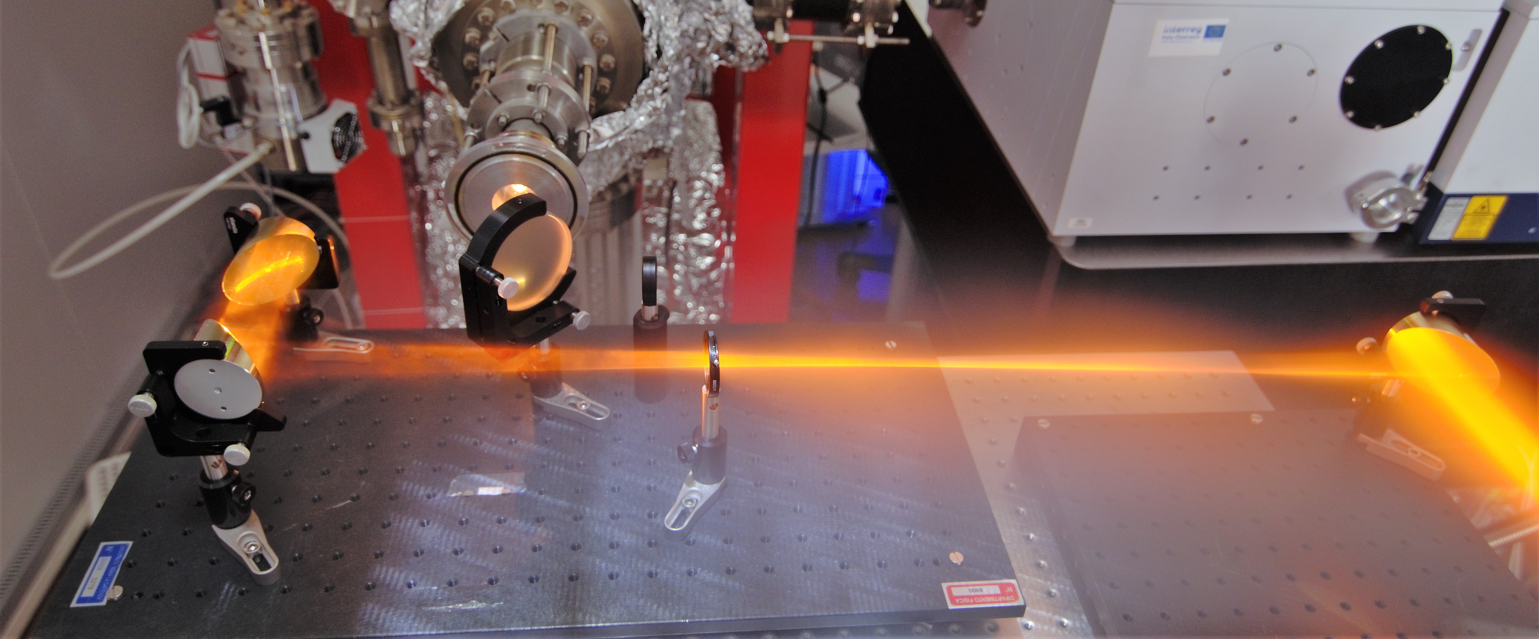
IMAGE PREVIEW
Figure 2: on-air path of the visible component of the IRSR of SISSI
Life Sciences and Plant Biology selected publications:
Contribution of Ribonucleic Acid (RNA) to the Fourier Transform Infrared (FTIR) Spectrum of Eukaryotic Cells. P. Zucchiatti, E. Mitri, S. Kenig, F. Billè, G. Kourousias, D.E. Bedolla, L. Vaccari. Anal. Chem. 88, 12090-12098 (2008)
Time-Resolved FT-IR Microspectroscopy of Protein Aggregation Induced by Heat-Shock in Live Cells. E. Mitri, S. Kenig, G. Coceano, D.E. Bedolla, M. Tormen, G. Grenci, L. Vaccari. Anal. Chem. 87, 3670–3677 (2015)
Multi-technique microscopy investigation on bacterial biofilm matrices: a study on Klebsiella pneumoniae clinical strains. G. Birarda, A. Delneri, C. Lagatolla, P. Parisse, P. Cescutti, L. Vaccari, R. Rizzo. Anal Bioanal Chem. 411, 7315-7325 (2019)
Environmental Science selected publications:
Plastics everywhere: first evidence of polystyrene fragments inside the common Antarctic collembolan Cryptopygus antarcticus. E. Bergami, E. Rota, T. Caruso, G. Birarda, L. Vaccari, I. Corsi. Biol. Lett.16, 20200093 (2020)
Cigarette butts, a threat for marine environments: Lessons from benthic foraminifera (Protista). F. Caridi, A. Sabbatini, G. Birarda, E. Costanzi, G. De Giudici, R. Galeazzi, D. Medas, G. Mobbili, M. Ricciutelli, M.L. Ruello, L. Vaccari, A. Negri. Mar. Environ. Res. 162, 105150 (2020)
Cultural Heritage selected publications:
The earliest evidence for mechanically delivered projectile weapons in Europe. K. Sano, S. Arrighi, C. Stani, D. Aureli, F. Boschin, I. Fiore, V. Spagnolo, S. Ricci, J. Crezzini, P. Boscato, M. Gala, A. Tagliacozzo, G. Birarda, L. Vaccari, A. Ronchitelli, A. Moroni, S. Benazzi. Nat Ecol Evol 3, 1409–1414 (2019)
Reflection FTIR spectroscopy for the study of historical bowed string instruments: Invasive and non-invasive approaches. G. Fiocco, C. Invernizzi, S. Grassi, P. Davit, M. Albano, T. Rovetta, C. Stani, L. Vaccari, M. Malagodi, M. Licchelli, M. Gulmini. Spectrochim. Acta A 245, 118926 (2021)
THE NANOSCOPY APPARATUS

IMAGE PREVIEW
Figure 4: sketch of the physical phenomenon at the basis of the nano-FTIR s-SNOM

IMAGE PREVIEW
Figure 5: nano-IR branch of SISSI
The nanoscopy apparatus installed at SISSI beamline is the neaSNOM microscope by Neaspec, based on an atomic force microscope (AFM). Thanks to the AFM functionality, the system allows reconstructing the nanoscale topography of the sample. In addition and more importantly, the neaSNOM microscope permits additionally nanoscale optical measurements.
The functionality presently implemented on the neaSNOM microscope set-up at SISSI beamline (branchline SISSI-Nano) is the nano-FTIR spectroscopy. The fundamental behind this functionality is the scattering-type scanning near-field optical microscopy (s-SNOM). In s-SNOM, the IR light is focused onto a metallic AFM tip, that confines the light under its tip apex in a 10nm small local hotspot. By raster scanning the tip onto the sample and detecting the elastically scattered light from the hotspot, an optical image with 10nm spatial resolution can be obtained. This method bypasses the ubiquitous diffraction limit of light and provides an unique optical access to the nanoworld. In nano-FTIR at SISSI, the IR light derives from a mid-IR source, a broadband laser based on fiber laser and difference frequency generation (DFG) or the ultra-broadband beam from the SISSI beamline (optical coupling is on-going). The elastically scattered light from the hotspot underneath the AFM tip is detected through an asymmetric interferometer and a spectrum is obtained via Fast-Fourier Transformation analogous to conventional FTIR-spectroscopy. This hence allows chemical identification of materials on the 10 nanometer length-scale. More precisely, s-SNOM provides information about the complex optical properties of a sample under the metallized tip: both the optical amplitude and phase of the scattered light can be measured. With appropriate models, the complex optical constants (n, k) of the material can be retrieved and the optical phase versus wavelength provides a good approximation to a conventional IR absorption spectrum. The s-SNOM technique works on a variety of materials, but the best signal to noise is obtained for materials with high reflectivity and high dielectric constants, namely for metals and semiconductors, but also metalorganic compounds such as pigments, or hybrid materials.
