FACILITY AT NFFA-TRIESTE
The JEOL™ JEM-2010F Ultrahigh Resolution TEM/STEM is installed in a dedicated laboratory at the CNR-IOM Institute and made available for access as part of the NFFA-Trieste project. It is built by anchoring a concrete platform directly onto the karst rock with only weak links to the laboratory.
The TEM microscope has a Thermally Assisted Field Emission Gun (FEG) electron source with a ZrO/W [100] filament. The high-brightness source produces a highly coherent electron probe with a diameter of less than 0.13 nm and a resolution limit of 0.11 nm in phase contrast. The small size of the probe produces sub-nanometer resolution in analytical and spectroscopic measurements.
The instrument is currently equipped with an 80mm2 Oxford X-Max Silicon Drift Detector for or Energy-Dispersive x-ray Spectroscopy analysis (EDS) allowing detection of light elements (Z > 5).
The available Scanning Transmission Electron Microscopy (STEM) attachment coupled with EDS can be used to obtain chemical profiles with high spatial resolution.
In addition, the coupling between the STEM attachment and the High-Angle Annular Dark Field (HAADF) detector is used to obtain Z-contrast images.
Sample preparation facility for (S)TEM
- Precision cutting tool for TEM sample preparation
- Disc grinder (Gatan, Inc) for TEM sample preparation
- Dimple grinder system for pre-thinning (Gatan, Inc)
- Precision ion polishing system (PIPS) (Gatan, Inc)
- Optical microscope
- Plasma cleaner
TEM Specimen Holders
- In-situ TEM holder
- Single/double tilt holder

IMAGE PREVIEW

IMAGE PREVIEW
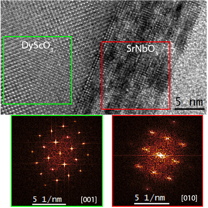
IMAGE PREVIEW

IMAGE PREVIEW

IMAGE PREVIEW

IMAGE PREVIEW
DFT calculated relaxed structure of La0.7Sr0.3MnO3

IMAGE PREVIEW
LSMO

IMAGE PREVIEW
Schematic model of LaAlO3 /Ti2AlO4 heterostructure
First-principle calculations
Experimental TEM observations are supported by atomistic simulations thanks to the CNR-IOM Democritos computing cluster, in particular density functional theory (DFT) calculations to more accurately determine the structure of the specimen and evaluate the structure-property correlations.
The Quantum Espresso open-source DFT-package allows for the interpretation of data often dealing with different types of defects, formation of a new phase at the interface, and analysis of grain-boundaries mainly in transition metal-oxide heterostructures.
Integration with other techniques
Precision Ion Polishing Systems (PIPS) enable high-quality TEM specimens to be obtained by gently milling at lower energies and at grazing incidence of solid-state materials.
All experiments are supported by image simulation and data analysis treatment of the results obtained both in imaging and diffraction mode.
The work is carried out in strong synergy with the growth and synchrotron-based electronic characterization NFFA-Trieste techniques: tipically, material systems are grown with PLD and MBE. Samples are first screened and investigated by SEM, then prepared in different geometries (plan-view and cross-section) by mechanical polishing and further explored at the atomic level by TEM/STEM to determine the relationships between structural, functional and electronic properties.

IMAGE PREVIEW
JEOL™ JEM-2010F Microscope

IMAGE PREVIEW

IMAGE PREVIEW

IMAGE PREVIEW
